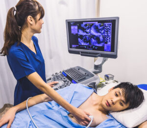
Eletrocardiography is the (transthoracic) ultrasound examination of the heart.
An echocardiograph involves bouncing sound waves off the heart to analyse structure. In order to answer the question “What Shape Is My Heart In?”, it gives an accurate picture of the size and strength of heart muscle, and the potency of the heart valves. Examples of investigation are:
- Examining heart muscle status, for instance, thickening ( hypertrophy) associated with high blood pressure; or weakening( dilatation) with cardiomyopathy, after a heart attack, or as an inherited condition;
- Assessing recovery from a heart attack and anylocalised injury, although this is unusual these days after intervention with a stent; and
- Assessing heart murmurs, after heart sounds are heard with a stethoscope; the type and severity of heart valve murmurs are diagnosed.
- Correlation of the image with the clinical picture is an area where a medical opinion can set a clear direction .

