Echocardiography

What is it?

Eletrocardiography is the (transthoracic) ultrasound examination of the heart.

An echocardiograph involves bouncing sound waves off the heart to analyse structure. In order to answer the question “What Shape Is My Heart In?”, it gives an accurate picture of the size and strength of heart muscle, and the potency of the heart valves. Examples of investigation are:

  • Examining heart muscle status, for instance, thickening ( hypertrophy) associated with high blood pressure; or weakening( dilatation) with cardiomyopathy, after a heart attack, or as an inherited condition;
  • Assessing recovery from a heart attack and anylocalised injury, although this is unusual these days after intervention with a stent; and
  • Assessing heart murmurs, after heart sounds are heard with a stethoscope; the type and severity of heart valve murmurs are diagnosed.
  • Correlation of the image with the clinical picture is an area where a medical opinion can set a clear direction .
Echocardiography

What should I expect?

Echocardiography is not dangerous and is usually painless, and does not involve radiation or X-rays. You do not need to specially prepare for an echocardiogram.

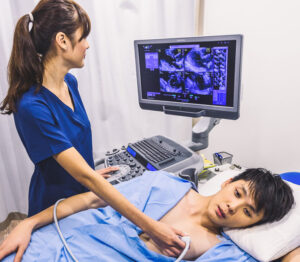
During the test, you will lie on an examination bed and the technician will put a thick gel on your chest. The gel may feel cold but it does not harm your skin. The technician then uses the transducer to send and receive inaudible sound waves.

The transducer will be placed directly on the left side of your chest, over your heart. The technician will press firmly and moves the transducer across your chest, and will ask you to briefly hold your breath during the test. For most of the test you will lie still.

Stress Echocardiography

What is it?

Stress echocardiography assesses the likelihood that you have significant narrowings in your heart arteries. By comparing your heart function before and after exercise using echocardiography, gives information which can your infer whether enough blood is getting to your heart or if a narrowing is restricting this supply.

What should I expect?

At the beginning of the test, your heart will be imaged using ultrasound (called echocardiography). The technician will put a thick gel on your chest, which may feel cold but does not harm your skin. Then, the technician will use the transducer to send and receive inaudible sound waves. The transducer will be placed directly on the left side of your chest, over your heart. The technician will press firmly as he or she moves the transducer across your chest and will ask you to briefly hold your breath during the test.

For an exercise stress echocardiogram, you will walk on a treadmill and the grade and speed will increase every 3 minutes.

This will make you feel like you are walking uphill. This increasing workload on the heart ensures the test is as accurate as possible. You should exercise for as long as possible to ensure your test is as accurate as it can be.

After you have finished exercising, you will lie down on an examination bed and the technician will take more echocardiography pictures. You will be asked to breathe out and hold your breath briefly several times so the images can be recorded. It is very important that you do your best to hold your breath during this part of the examination, because the technician has only 1-2 minutes to obtain these images before your heart slows down to its normal rate.

Complications are rare during both exercise stress echocardiography. Heart attack or abnormal heart rhythms may occur rarely, but the risk of death is about 1 in 10,000.

Duration is one hour, and preparation : nil . Please bring previous results with you